Using innovative research approaches involving genetics and stem cells, UT scientists are attempting to solve the problem of long-term use of anti-diabetic drugs that appear to lead to weaker bones in some people who use them.

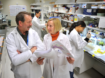
Dr. Beata Lecka-Czernik, right, confered with research aide Piotr J. Czernik as Vipula Petluru, predoctoral research assistant, right, and Dr. Shilong Huang, postdoctoral fellow, conducted experiments.
Dr. Beata Lecka-Czernik is directing the basic-science studies aimed at coming up with ways to reduce the effects of a new class of anti-diabetic drugs known as TZDs, which appear to adversely affect bone health. The best known of these drugs are Avandia and Actos, which work by sensitizing the body to insulin and are considered breakthrough medications for blood-sugar control. They were introduced in the late 1990s.
That is good news for the estimated 16 million Americans who have type 2 diabetes, a condition in which the body does not properly use insulin.
Her studies, which are funded with a $1.5 million grant from the National Institutes of Health and a $300,000 grant from the American Diabetes Association, rely on the fact that the human body’s 206 bones are in a constant state of remodeling — dissolving small bits of old bone, a process called resorption, and rebuilding new bone. Good bone health is a combination of bone formation and bone resorption. As people age, bone loss exceeds production, leading to osteoporosis.
Bone cells, which are called osteoblasts, and fat cells evolve from the same kind of stem cells called mesenchymal cells that are found in tissue inside the bone cavity.
However, in a 2004 study reported in the journal Endocrinology, which garnered national publicity, Lecka-Czernik demonstrated in mouse studies that bone-marrow stem cells are more likely to become fat cells, increasing the fat content in bone instead of forming osteoblasts when a signaling protein called peroxisome proliferator-activated receptor-gamma (PPAR-gamma) is activated by anti-diabetic TZD drugs. As a result, mice that received the drugs lost bone and developed osteoporosis.
Her study was followed by several clinical studies demonstrating that people, especially older women, who take TZDs for at least one year were more likely to lose bone and have a broken bone than those who did not take TZDs, Lecka-Czernik said.
So while diabetics may find the insulin-stimulating drug extremely effective in controlling blood sugar, it may put them at higher risk for bone loss, according to the UT researcher.
“This is not an acute effect, but an accumulative effect,” she explained. “These are very valuable drugs that have to be taken for a lifetime. Although they don’t cure diabetes, they very effectively control blood-sugar levels. So we know that with longer use of these drugs, we will see greater bone loss and an increase in human bone fractures. We are afraid that over time diabetic patients on TZDs will become a group at risk for orthopedic complications, including frequent bone fractures and healing complications.”
Lecka-Czernik, one of the country’s leading authorities on changes that occur to bone during aging and diabetes, and her Health Science Campus research team — Drs.Shilong Huang, Meenakshi Kaw and Piotr Czernik and graduate students Sima Rahman and Vipula Petluru — plan to take several experimental approaches to increase the safety of TZD drugs on bone, protect against bone loss, and improve fracture healing in patients on TZD therapy.
One approach uses mice to test which FDA-approved therapies work best to protect against TZD-induced bone loss. They include teriparatide injection to induce bone formation and bisphosphonate administration to inhibit bone resorption.
To analyze the bone-repair process, the researchers have developed sophisticated technology to first drill a tiny hole in the tibia of anesthesized mice. In Petri dishes containing nutrients, vitamins and growth factors, stem cells will be grown with a gene that coaxes them to become bone-forming cells called osteoblasts. The researchers then inject the stem cells into the fracture site.
“We will experiment with the methods of increasing bone formation by using genetically modified stem cells,” Lecka-Czernik explained. “This therapy is experimental and not yet approved for treatment of humans. Genetic modification means that we will either introduce into these cells a gene that is beneficial for bone formation, or we will inhibit a gene that has a negative effect on this process. Basically, we will manipulate these cells in the Petri dish and than inject them into a fracture site to induce bone healing. After that, we plan to conduct experiments to manipulate stem cells directly in the bone, at the fracture site, without a need to manipulate them outside the bone.”
The scientists use sophisticated imaging techniques called micro-computed tomography to analyze the bones of the mice for new bone formation.
Lecka-Czernik previously was a faculty member at the University of Arkansas for Medical Sciences and started duties at UT in 2007 as a professor in the departments of Orthopedic Surgery and Physiology/Pharmacology. Her studies are being conducted at UT’s Center for Diabetes and Endocrine Research on Health Science Campus.
A member of several scientific journal editorial boards and an NIH grant reviewer, Lecka-Czernik has been invited to serve as a member of the organizing committee and a speaker for the next topical meeting of the American Society for Bone and Mineral Research that is titled “New Frontiers in Skeletal Research: Bone, Fat and Brain Connections.” It will be held April 27-28 in Bethesda, Md.