The common mouse has a pretty special characteristic.
The sperm cells of mice, along with genetically similar species in the family Muridae, don’t have recognizable centrioles. While these subcellular organelles are essential for embryo development in most animals, mice don’t need them. They can do the job with just the nucleus of a sperm cell.

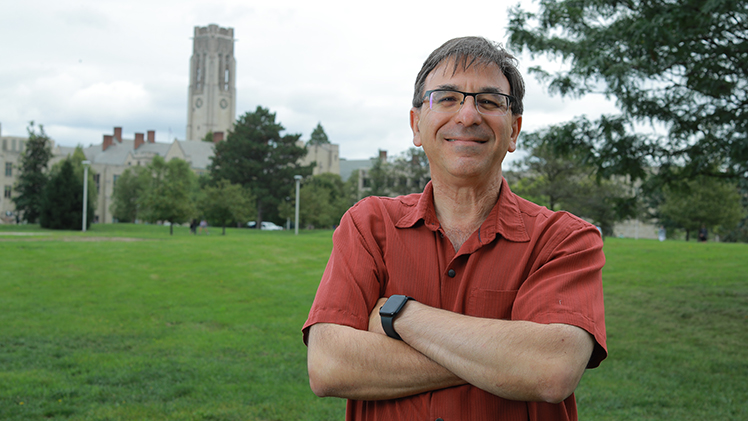
Dr. Tomer Avidor-Reiss, a professor in the Department of Biological Sciences, explores an unusual mechanism in mouse reproduction in new research published in Nature Communications.
It’s a trick humans, bovines and other species don’t have – and in some cases would very much like.
“It would solve a lot of infertility cases if you were able to fertilize an egg with just the nucleus of a sperm cell,” said Dr. Tomer Avidor-Reiss, a professor in the Department of Biological Sciences.
Avidor-Reiss explores this mechanism in research published this month in Nature Communications. His article, titled “The Evolution of Centriole Degradation in Mouse Sperm,” proposes an evolutionary explanation for the absence of centrioles in house mice, rats and similar species that he believes scientists could build on as they continue to advance human reproductive science.
“That’s our motivation,” Avidor-Reiss said. “Can mice tell us something that will be useful in helping people whose centrioles are abnormal, and who are struggling with infertility as a result?”
The research is the latest of several publications to come out of his lab at The University of Toledo, much of it building directly on ground-breaking research he published in Nature Communications in 2018. That article proved the existence of a previously unknown second centriole in human and most other mammalian sperm cells, atypical in shape compared to the long-identified canonical centriole.
Students in an undergraduate biology research lab shared a byline in microPublication Biology in late September, exploring the localization of centriole proteins in cattle and human spermatozoa. This is the second class-written paper to be published under the guidance of Avidor-Reiss.
Abigail Royfman, a current medical student and former undergraduate researcher in his lab, is the lead researcher on a book chapter titled “Structural Analysis of Sperm Centrioles Using N-STORM,” referring to a new super-resolution microscopy. This chapter was published online in October.
And Avidor-Reiss and his collaborators published a research article in October in Scientific Reports, another journal published under the Nature Portfolio. Leading this research were doctoral candidates Katerina Turner and Luke Achinger, joined by a past doctoral candidate, Dr. Emily Lillian Fishman, and undergraduates Derek Kluczynski and Audrey Phillips. They also collaborated with Dr. Barbara Saltzman of the UToledo Department of Population Health, Dr. Bo Harstine of Select Sires and Dr. Dong Kong and Dr. Jadranka Loncarek of the National Cancer Institute.
In this article they proved they can identify reduced fertility in bulls using a patent-pending lab test that identifies abnormalities in centrioles they developed at UToledo.
The researcher explained that reduced fertility, also called subfertility, is a more subtle parameter than infertility. It refers to a difficulty, rather than an inability, to conceive.
“Subfertility matters in the world of agriculture and artificial insemination, where a rate of conception that’s even slightly lower than the average can have a multiplier effect across thousands of cows,” Avidor-Reiss said. “Artificial insemination companies put their bulls through an extensive array of semen analyses because they want to market high-quality sperm to farmers. For them it’s reputation, it’s business. Our research is significant because we show that our lab test can not only identify infertility, not only identify infertility that’s not explained by standard tests, but also subfertility.”
His and his collaborators’ most recent research, in Nature Communications, digs into a long-accepted enigma when it comes to the study of centrioles: Why do mice, rats and similar species seemingly have none at all?
Avidor-Reiss and his collaborators begin their article by challenging that conclusion. They point to what they call “remnant” centrioles in murid sperm cells that contain the expected proteins, just not in the expected location or structure within the cell.
They also propose an explanation: Mice ancestors once had identifiable centrioles like other rodents, but underwent an evolutionary process through which these organelles became dispensable to embryo development. That instigated their degradation to their current form.
Avidor-Reiss explained that the research is significant because it is the first molecular evidence of the rapid diversification and adaptive evolution of centrioles.
It also presents a launching point for future research, he said.
“This is the first step to really get into the mechanism of how mice can create life without sperm centrioles,” he said. “They are unique in that. Other mammals cannot do it.”