Most scientists focus on what’s under their microscope.
Dr. Aniruddha Ray tends to concern himself with the instrument itself.
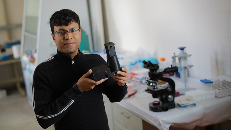
Dr. Aniruddha Ray, an assistant professor in the Department of Physics and Astronomy, is developing a low-cost, high-resolution model intended to significantly advance the development of anti-cancer drugs
He’s developing a low-cost, high-resolution model intended to significantly advance the development of anti-cancer drugs, which is currently hindered by a limited availability of high-tech, high-cost microscopes. His work is supported by a $451,500 grant through the National Institute of Biomedical Imaging and Bioengineering.
“If we can spend $100 on a microscope instead of $100,000, you can imagine how paradigm-shifting that would be,” said Ray, an assistant professor in the Department of Physics and Astronomy. “It’s essentially democratizing this technology.”
Ray is collaborating on this research project with Dr. Devinder Kaur, a professor in the Department of Electrical Engineering and Computer Science, and Dr. Amit Tiwari, associate dean of research and graduate studies at the University of Arkansas for Medical Sciences College of Pharmacy and a former professor in UToledo’s Department of Pharmacology and Experimental Therapeutics.
The anti-cancer drugs they’re eyeing through this research are alternatives to common treatments like chemotherapy, which work by inducing programmed cell death or apoptosis. Scientists have found that multidrug resistant cancers are highly resistant to treatments that rely on this mechanism, presenting a major public health concern and an incentive to develop non-apoptotic therapies.
These promising therapies have proved difficult to develop, though. Biochemical assays and high-definition imaging are the only methods for studying them, and each require significant time, expense and skill, which limits their utility to a small number of laboratories.
Enter Ray, whose expertise is in biophotonics, a fast-growing field that applies light-based technologies to problems in the life sciences and medicine. His proposal uses photonics principles to bring down the cost of critical microscopes without bringing down their quality.
How?
“It’s in how we design both the hardware and the software,” Ray explained.
His proposal is leagues away from the standard optical microscopes that line high school classrooms by the dozens – and even from their much higher-powered counterparts in prestigious research labs that are currently being used in anti-cancer drug research. It calls for a lens-less holographic microscope, which uses LED light sources, off-the-shelf optical components and an image sensor to create holograms that a computer then processes to produce images in a three-dimensional plane.
One benefit of forgoing the lens is that it doesn’t sacrifice the field of view for resolution, as is the case with conventional optical microscopes that must zoom in on a sample to obtain a more detailed image.
“We’re bypassing that,” Ray said. “When you’re studying cancer cells, you need to observe a lot of them. You can’t just say, well, this new drug worked on five cells. You have to see it work on thousands. With the microscope design that we use, you can image thousands of cancer cells at one time.”
That’s the hardware. When it comes to the software, Ray is turning to artificial intelligence to analyze the resulting mountains of images and turn all that raw data into something researchers can use.
“We don’t need to sit there for hours and days, looking at these images and trying to figure out what’s happening,” he said. “We don’t need specially trained experts to do that. You just put the sample in, it gets automatically imaged and then this artificial intelligence program that analyzes the data provides the answer, almost in real time.”
While he and his collaborators will be focusing the high-tech tool they develop on cancer cells for now, Ray anticipates the impact of a low-cost, high-resolution microscope will extend beyond this line of research.
“If this is successful, it could reduce the amount of time it takes to bring a drug to the market by several years,” he said. “A lot of labs that work on drug discovery processes would have access to instruments they can afford. They wouldn’t have to rely on expensive microscopes, or send their samples to another lab and then wait for results to come back.”